EPI Education
Take a deep dive into EPI and learn from experts in the field.
About EPI: EPI pathogenesis, symptoms, and diagnosis
Etiologies of EPI: Recognize EPI and the barriers to diagnosis
Management of EPI: Key information for managing EPI
Case Studies: Experts present case-based narratives on EPI
Select one or more filters below to explore topic-specific resources.
Transcript
This is a women, 62 years old, presenting with loose stools over the last year, characterized by two bowel movements, of foul smelling, sometimes, not always, sometimes greasy or oily large volume floating stools. Frequently postprandial, but occasionally nocturnal, no urgency or incontinence, and no signs of inflammatory diarrhea. As additional symptoms, she refer abdominal pain. And you know, that lady, because of the presence of pain, abdominal pain and diarrhea, was referred to us from pi- primary care doctor with the diagnosis of IBSD. But when we asked her if the pain was not really related to bowel movements, she referred also bloating and flatulence, unintentional weight loss of three, four kilograms, over the last year, not so amazing. And she was not a drinker, smoker of 10 cigarettes per day. She was previously diagnosed with type 2 diabetes just six months before she was on metformin, and this is something important to take into account, because you know that metformin is one of the drugs causing diarrhea. The family history was not remarkable in terms of digestive diseases.
So, the general examination was unremarkable except for epigastric tenderness to deep palpation, and the lab test that I told you before were normal. So the blood cell count was normal, the metabolic pa- panel was normal, celiac serology was negative, fecal calprotectin was negative, and gly- uh glycemia, and HbA1C was markedly high as a sign of poorly controlled diabetes. At that time, our differential diagnosis was, well, it can be a functional disorder, or a disease leading to maldigestion, and/or malabsorption of nutrients.
And to make this differential diagnosis we underwent an abdominal ultrasound, that was basically normal except because the pancreas was obscured by gas, which is quite frequent during abdominal ultrasound, upper GI endoscopy with duodenal biopsies were negative, lactose blood test was negative, and we also tested for fecal elastase [inaudible] pancreatic disease, and fecal elastase was markedly low, 58 microgram per gram. You know that normally is higher than 200 micrograms per gram. Because of that, we wanted to have a look into the pancreas, and we performed a CT scan, and as you can see on your right, CT scan shows a chronic calcifying pancreatitis.
And that was the final diagnosis. Chronic calcifying pancreatitis, with exocrine pancreatic insufficiency, and pancreatogenic diabetes. Not type 2 diabetes, but pancreatogenic diabetes. As the therapy, we recommend the smoking a- abstinence, non-opioid analgesics on demand for pain, pancreatic enzyme replacement therapy for EPI, and insulin for diabetes. Three months later, the patient was fully symptom free.

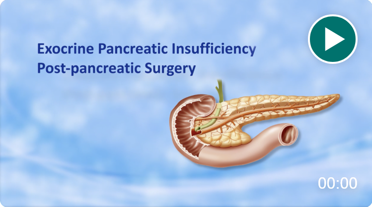
Exocrine Pancreatic Insufficiency Case Study
See how Dr. Domínguez-Muñoz evaluated a patient's symptoms and history to determine a diagnosis.

Take an immersive journey into the physiology of the exocrine pancreas and the pathophysiology of EPI.


